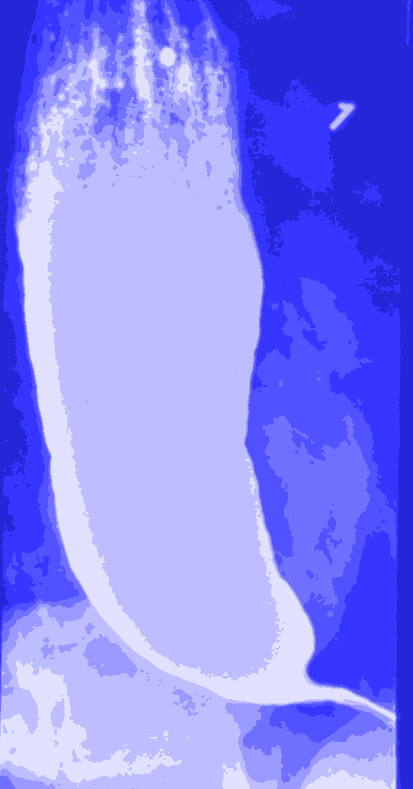
Histopathologic patterns among achalasia subtypes
- Dr. Steven Horwitz

- Nov 11, 2015
- 1 min read
Biopsies were taken from 46 myotomy patients: 20 type 1, 20 type 2, 3 type 3 and 3 EGJ obstruction.
The study suggests that Type 1 is a progression from type 2.
"On histopathology, complete ganglion cell loss occurred in 74% of specimens, inflammation in 17%, fibrosis in 11%, and muscle atrophy in 2%."
This means that there was complete nerve cell destruction in 74%, inflammatory changes in 17%, fibrosis or hardening of the muscle tissue lining of the esophagus in 11% and muscle atrophy (muscle cells smaller) in 2%.
19 of 20 type 1 had complete nerve cell destruction whereas it was 13/20 in type 2.
Interestingly, 3 patients had "normal" tissue: 1 type 2, 1 type 3 and 1 obstruction.
As I have said before, I think type 3 is just an earlier stage which eventually leads to type 1.


Comments